


University Research with 3D Optical Scanning
An interdisciplinary team of researchers was awarded $2.2 million from the National Institutes of Health to determine whether 3D optical scans could show a direct correlation between body shape and liver health.
An interdisciplinary team of George Washington University researchers received a $2.2 million grant from the National Institutes of Health for a project that combines 3D body surface scanning technology and machine learning to develop an inexpensive and noninvasive way to assess the health of highly obese individuals.
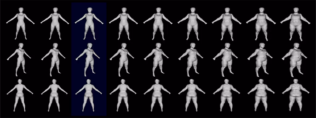
This new project seeks to use 3D depth cameras—special cameras that measure distances to the subject as well as the color for each pixel—to capture an individual’s body shape and correlate this to health indicators associated with obesity using machine learning algorithms. The team is focused specifically on fibrosis and a fat build up in the liver known as fatty liver disease, or hepatic steatosis. Fatty liver disease can eventually lead to liver failure and is currently diagnosed with an invasive biopsy.
“As they lose weight, we can then see what the relationship is between body shape and X, where X could be things like liver health or the percentage of body fat, or changes in their blood tests that are performed periodically throughout their recovery process,” Professor James Hahn said.
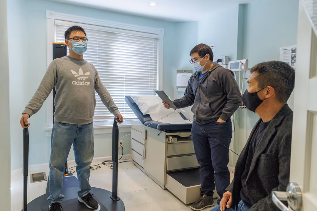
Hahn and the team will capture the body shapes of patients enrolled in the study using 3D depth cameras installed at the GW Weight Loss and Surgery Center. The patients will also undergo medical imaging at the Milken Institute School of Public Health. The school’s dual-energy X-ray absorptiometry, or DEXA for short, measures bone density and body composition like body fat and muscle mass. These imaging results, combined with other clinical tests and a liver biopsy taken before bariatric surgery will serve as the training dataset for the machine learning algorithm. Patients will return four times for optical body surface scans and DEXA imaging in the year following surgery, enabling researchers to track how their body shape changes as a function of health indicators over time.
Hahn’s team plans to compile this dataset and, using two sub-approaches in machine learning, train systems able to predict health indicators based on body shape, including hepatic steatosis, he said. The ability to draw this correlation has the potential to be less expensive, less invasive and/or more sensitive than existing diagnostic methods, Hahn said.
“This whole approach has a lot of potential,” Hahn said. “This technology is not going to replace biopsies or other medical tests, but it will perhaps give physicians a new cheaper and more convenient imaging modality they can employ for diagnosis.”
Collaborator and bariatric surgeon Khashayar Vaziri, a professor of surgery at SMHS and program director of general surgery residency at GW Hospital, said this approach could help physicians diagnose liver problems sooner.
“Bariatric surgery patients are just a small subset of the obese population. This would allow health care providers to apply this technology and get a better understanding and diagnose liver disease in this patient population much, much earlier,” Vaziri said. “Diagnosis of liver disease is elusive, and this would be a very useful tool.”
Hahn described the 3D body surface scanning the team uses as similar to the depth camera technology available on newer smartphones. Inexpensive compared to most medical imaging, this type of scan could be done in a regular clinic or doctor’s office or even at home.
This project builds on more than a decade of research in digital health. Past projects include a collaboration with USA Swimming prior to the 2008 Beijing Olympics to examine how elite swimmers could improve their strokes. Hahn created a 3D virtual representation of an Olympic swimmer and compared the fluid dynamics of the athlete to a dolphin—capturing body shape and using that data for different types of motion. Over time, he sought to identify how to capture body shape and motion simultaneously.
Original Article written by Kristen Mitchell at GW Today.


.png)
Subscribe to
our Newsletter!
